Ultrasound in Obstetrics and Gynaecology
Safe, Accurate, and Essential for Women’s Health
Ultrasound is a safe and painless imaging test that uses sound waves to create real-time pictures of internal organs and the developing baby during pregnancy.
It is one of the most widely used tools in women’s health, helping doctors monitor pregnancy progress, assess reproductive organs, and detect any problems early.
Ultrasound does not use radiation, making it completely safe for both mother and baby.
What Is an Ultrasound?
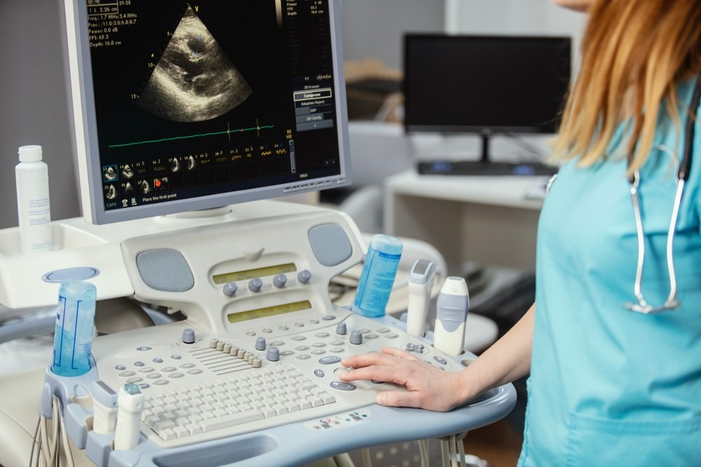
An ultrasound (or sonogram) uses high-frequency sound waves sent through a small handheld device called a transducer.
The images are displayed instantly on a monitor, helping doctors see the uterus, ovaries, and fetus clearly.
There are two main types:
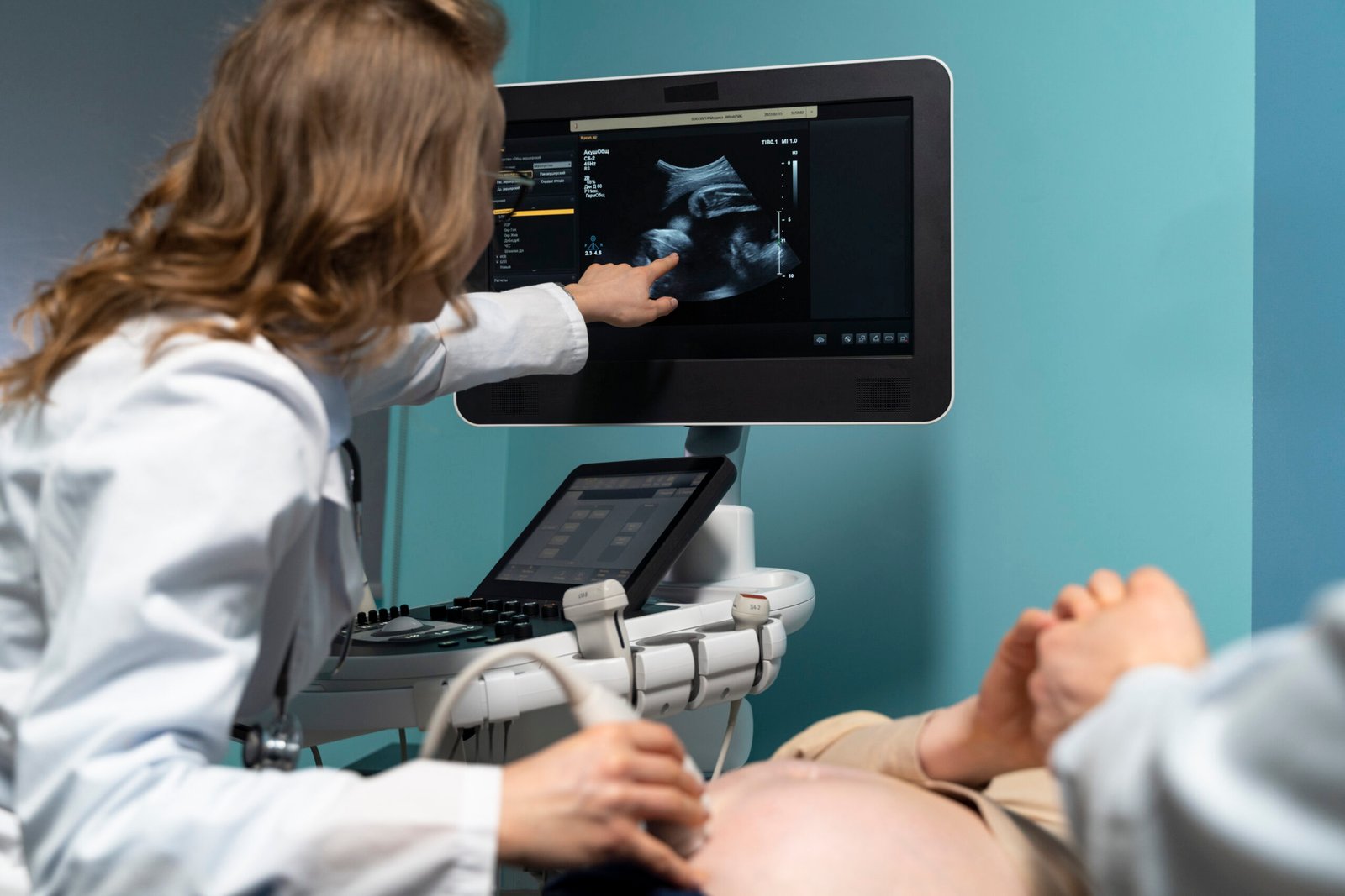
- Transabdominal Ultrasound: The probe is placed on the abdomen (with a full bladder).
- Transvaginal Ultrasound: A thin probe is gently inserted into the vagina for clearer internal images (usually done when early pregnancy or pelvic details are needed).
Both are safe, comfortable, and quick procedures.
Ultrasound in Obstetrics (During Pregnancy)
Ultrasound plays a key role in every stage of pregnancy — from confirming conception to checking your baby’s growth before delivery.
Early Pregnancy Scan (6–10 weeks)
- Confirms the baby’s heartbeat and position
- Detects single or multiple pregnancies
- Identifies early complications like ectopic pregnancy
Nuchal Translucency (NT) Scan (11–13 weeks)
- Measures fluid behind the baby’s neck to assess risk for chromosomal abnormalities (like Down syndrome)
- Often combined with maternal blood tests for screening
Anomaly Scan (18–22 weeks)
- A detailed scan that checks the baby’s organs, spine, limbs, and placenta
- Detects most major structural or developmental conditions
Growth and Doppler Scans (after 28 weeks)
- Monitor the baby’s growth, blood flow, and movement
- Assess placenta, cord, and amniotic fluid
- Detect early signs of fetal distress or growth restriction
3D / 4D Ultrasound
- Provides clear 3D or moving 4D images of your baby’s face and movements
- Enhances bonding and allows better visualization for clinical evaluation
Ultrasound in Gynaecology
Ultrasound is equally important in the diagnosis and management of various gynaecological conditions. It helps assess the uterus, ovaries, fallopian tubes, and surrounding pelvic structures.
Transvaginal Scan (TVS)
- Offers detailed images of the uterus, ovaries, and endometrial lining
- Commonly used to investigate irregular bleeding, pelvic pain, or infertility
Follicular Monitoring Scan
- Tracks egg development during fertility treatment or natural cycles
- Helps identify the best time for ovulation and conception
Saline Infusion Sonography (SIS)
- A small amount of sterile fluid is placed inside the uterus for a clearer view of the uterine cavity
- Helps detect fibroids, endometrial polyps, and adhesions
Pelvic Scan
- Evaluates overall pelvic health
- Detects ovarian cysts, fibroids, endometriosis, and other structural problems
Endometrial Thickness Assessment
- Measures the uterine lining in women with infertility, PCOS, or menopausal bleeding
- Helps guide treatment and assess hormonal balance
IUD or Implant Position Scan
- Checks the correct placement of intrauterine devices (copper T, Mirena, etc.)
- Ensures safety and effectiveness of contraception
Benefits of Ultrasound